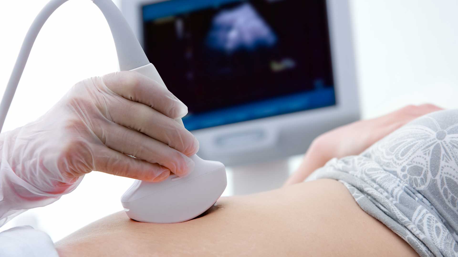
What is it?
CDS is recognised worldwide for our pioneering work in key areas of reproductive medicine and fertility.
We are able to offer our patients very advanced ultrasound imaging technologies, including:
- 2D and 3D transvaginal (internal) scanning
- Colour Doppler (blood flow) imaging
- Gynaecological procedures
- Virtual 3D ultrasound imaging
What do they show?
2D and 3D transvaginal (internal) scanning (TVS) have revolutionised and had a tremendous impact in the investigation and management of infertility. Advanced ultrasound imaging can identify gynaecological problems which may affect fertility, or normal development of early pregnancy. Gynaecological disorders might include the presence of endometriosis, fibroids, disorders of the ovaries or anatomical abnormalities.
Colour Doppler (blood flow) imaging: Modern colour Doppler imaging shows very fine blood flow through organ tissues. This allows adenomyosis (endometriosis within the muscle wall of the uterus) to be identified more clearly. It can also show blood flow changes within the ovary which reflect hormonal activity and ovulatory changes without the need for blood tests. Crucially, it also demonstrates the hormonal activity of the ovary essential for the development of early pregnancy.
Gynaecological procedures:
Enhanced ultrasound examination of the Fallopian tubes (HSS/HyCoSy) and uterine cavity (SIS) shows their overall healthiness.
Using ultrasound as guidance is a very effective and safe technique for biopsy of the lining of the womb. This is of particular value in the investigation of implantation problems.
Virtual 3D ultrasound imaging: CDS has collaborated with Canon Medical Systems to create the very first virtual 3D imaging system in gynaecology, as part of the SIS procedure. It produces very detailed life-like pictures of the inside of the womb cavity. This is immensely valuable in assessing the healthiness of the womb lining, or identifying anomalies within the womb cavity which could affect fertility or cause miscarriage.
For general enquires and further information please see our Contact Page

